
neck muscles anatomy
Use the mouse scroll wheel to move the images up and down alternatively use the tiny arrows on both side of the image to move the images on both side of the image to move the images. Longus Colli Capitis Responsible for flexion of the head and neck.
Pin By Firelight Healing Arts On Dentistry And Medicine Neck Muscle Anatomy Muscle Anatomy Shoulder Muscle Anatomy
Neck muscles are bodies of tissue that produce motion in the neck when stimulated.

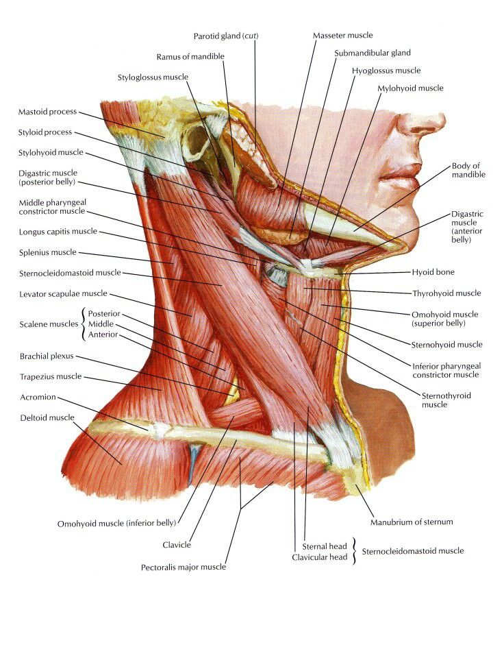
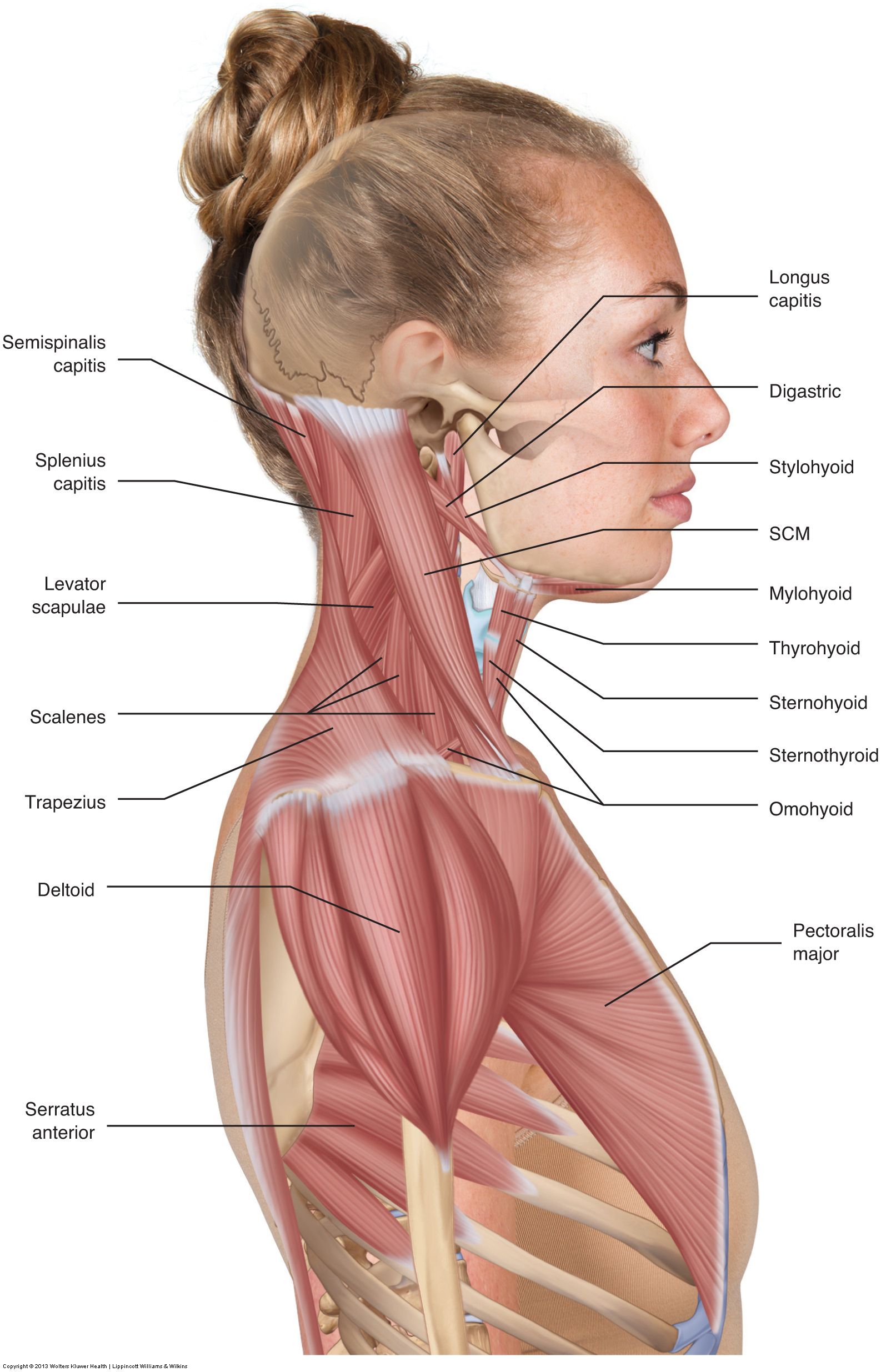
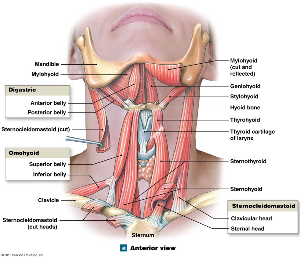
. The muscles of the neck are present in four main groups. This group of muscles comprises the sternohyoid omohyoid thyrohyoid and sternothyroid muscles. This MRI neck axial cross sectional anatomy tool is absolutely free to use. From the back they begin just beneath the base of your skull and extend down near the middle of your back around your shoulder blades.
From the front these muscles begin at your jaw and extend to your collarbone at the top of your chest. Watch Cervical Muscle Anatomy Animation. In the upper posterior part of the neck below the occipital bone the four paired suboccipital muscles. The neck is one of the most complex and intricate structures in our body and includes the spinal cord which sends messages from the brain to the rest of the body.
Suboccipital muscles Suprahyoid muscles Infrahyoid muscles Scalene muscles These four muscles are described below. They move the larynx and depress the mandible. The levator scapulae muscle is attached at the top four cervical vertebrae C1 to C4 and runs down the. They are responsible for head movement stabilizing the upper region of the body assisting in swallowing helping to elevate the rib cage.
It covers the frontal and lateral side. Its submitted by management in the best field. Causes of Neck Pain and How to Manage the Pain In basic terms the neck cervical spine joins the shoulders and chest to the head. Your neck muscles are amazing.
They are supplied segmentally from Cl 2 and 3 via the ansa cervicalis. Many medical conditions affect these muscles and an impair their structure as well as their function. The muscles of the neck are closely related to a number of important structures that pass between the thorax and the head including major blood vessels nerves and elements of the respiratory and gastrointestinal systems. They are usually described within the triangles.
Deep Neck Muscle Anatomy. Flexible Online Learning at Your Own Pace. So there are the muscles of the anterior triangle and the muscles of the posterior triangle. Hypaxial muscles generally lie ventral to the transverse processes of the vertebrae.
Ad Build your Career in Data Science Web Development Marketing More. Your neck muscles are at the front back and sides of your neck. There are many muscles around the neck that help to support the cervical spine and allow you to move your head in different directions. Some also create.
This article will introduce you to the anatomy of the muscles of the neck. We understand this kind of Deep Neck Muscle Anatomy graphic could possibly be the most trending subject subsequently we ration it in google gain or. Neck muscles help support the cervical spine and contribute to movements of the head neck upper back and shoulders. Neck Anatomy Muscles Pictures.
The suprahyoid muscles are a group of neck muscles located above the hyoid bone and all elevate this bone while the infrahyoid muscles are situated below the hyoid bone and participate in depressing it. Epaxial muscles generally lie dorsal to the transverse processes of the vertebrae and are associated with the vertebral column and ribs. Neck Muscle Anatomy A look at the Sternocleidomastoid and Scalenes. The mimetic muscles are considered to be an extension of the superficial musculoaponeurotic system SMAS of the face which is a fascial plane deep to the subcutaneous tissue but superficial to the muscles of mastication running from the platysma in the neck up to the galea aponeurotica and the temporoparietal fascia under the scalp.
As the suprahyoid and infrahyoid muscles. Furthermore the anterior triangle muscles are grouped depending on their position to the hyoid bone. Here are some of the key muscles attached to the cervical spine. Rectus Capitis Anterior Responsible for.
They move the head in every direction pulling the skull and jaw. Anatomy of The Neck. 31 Cross section of the main muscles and fascial layers of the neck 32 Muscles of the Neck 321 Platysma Compared to all other muscles of the neck which are skeletal this is the only cutaneous. It is a wide and thin lamina in the subcutaneous tissue.
Superficial muscles are the muscles closest to the skin surface and can usually be seen while a body is performing actions. Neck muscles are of four types. Arteries Brachiocephalic left Common Carotid left External Carotid Internal Carotid Subclavian left. The suboccipital muscles act to rotate the head and extend the neckRectus capitis posterior major and Rectus capitis posterior minor attach the inferior nuchal line of the occiput to the C2 and C1 vertebrae respectivelyObliquus capitis superior also extends from the occiput to C1 while obliquus capitis inferior originates.
Invest 2-3 Hours A Week Advance Your Career. The neck muscle anatomy can further be divided into anterior lateral and posterior triangles. Suboccipital Muscles The suboccipital muscles are a group of four muscles that located under the occipital bone. The neck muscles including the sternocleidomastoid and the trapezius are responsible for the gross motor movement in the muscular system of the head and neck.
The prevertebral muscles of the neck are situated anterior to the spine. In this chapter we will dissect some hypaxial muscles associated with the neck thorax and abdomen. To perform clinical neck treatment that is accurate and specific the therapist needs to know the attachments and actions of the muscles of the neckFor example to know where to place the palpating hand when applying deep tissue work into the neck the manual therapist must know the attachments of the target muscle to be worked to be able to locate. Many in the neck help to stabilize or move the head.
These muscles are located in the sternocleidomastoid Trapezius splenius and. The muscles of the neck are a hot topic within anatomy circles. Later in this chapter we will dissect some of these muscles that. The strap muscles are retracted to access the trachea and thyroid gland and also form the anterior boundaries of the neck levels.
Anatomy Where are the neck muscles located. Here is a list of the many muscles that exist in the neck. Here are a number of highest rated Deep Neck Muscle Anatomy pictures upon internet. We identified it from obedient source.
The muscles of the neck run from the base of the skull to the upper back and work together to bend the head and.

Neck Muscle S And Nerve S Neck Muscles And Nerves Neck Muscle Anatomy Health Medicine And Neck Muscle Anatomy Muscle Anatomy Shoulder Muscle Anatomy
Human Body Anatomy Human Muscle Anatomy Body Anatomy
Pin On Career

Neck Muscle Anatomy Neck Muscle Anatomy Sternocleidomastoid Muscle Muscle Anatomy

The Human Muscle System Neck Muscle Anatomy Muscles Of The Neck Muscle Anatomy
Post a Comment for "neck muscles anatomy"